It's like a CT scan, but for an individual cell!

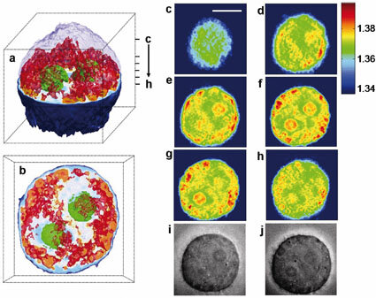 a. A 3D rendered image of a HeLa cell. The outermost layer of the upper hemisphere of the cell is omitted to visualize the inner structure. Nucleoli are colored green and parts of cytoplasm with refractive index higher than 1.36 are colored red. b. Top view of a.[via China View]
BEIJING, Aug. 14 (Xinhuanet) -- An imaging technique that could set a new standard in dozens of fields from immunology to neurology allows scientists to scan a living cell and render it as a 3-D image in a process similar to CT scanning now used in health care."Accomplishing this has been my dream and a goal of our laboratory for several years," Michael Feld, director of MIT's Spectroscopy Laboratory, told LiveScience.Until now, techniques for rendering cells in 3-D required the application of chemicals and stains, freezing and other invasive processes. These techniques interfere with normal cellular function to varying degrees, but that has not stopped their widespread use.The new technology can be used on live cells in their native state, with no preparation.Developing this process required that the scientists look to other fields that depend heavily on 3-D imaging techniques such as paleontology where CT scans are used to study fragile bones.The scan collects several narrow X-ray cross sections, or slices, of a 3-D object. The cross sections depict the density highs and lows of one thin section. Many slices are collected from several different orientations and then stitched together into a contiguous solid, much like making a loaf of bread out of individual slices.The MIT researchers used visible light instead of X-rays, but had to compensate for the fact that cells absorb very little light. To compose the images, they had to measure how much the light waves passing through the cell slowed, a property known as refractive index.After taking 100 slices measuring the cell's refractive index, the researchers composed a 3-D map that detailed the cell's many parts, from membrane to mitochondria."This will open up the possibility of imaging through tissues, which will have a significant impact on life science," said Wonshik Choi, first author of the study describing the technique. The study is published in the Aug. 12 online edition of Nature Methods.